Patellar luxation
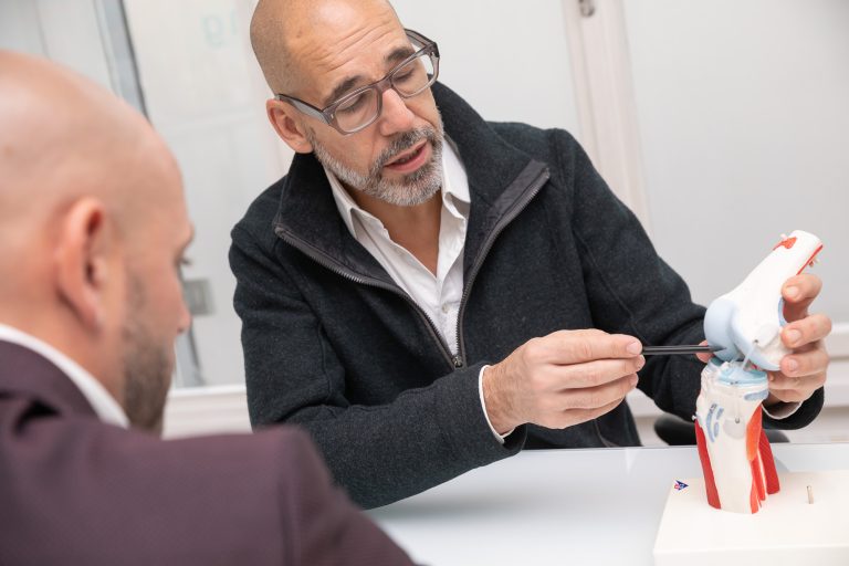
Treatment at the orthopedist from Vienna
With the kneecap, it’s all a question of the stability of the joint. Only when the kneecap functions properly can we extend and bend our knee without pain.
The kneecap, or patella, refers to a flat, disc-shaped bone that lies in front of the joint of the knee, in whose articular surfaces it participates and thus protects it. It is embedded in the large extensor tendon of the hamstring muscle and thus functions as the lever arm of the muscle.
OVERVIEW
The patella in a patellar dislocation
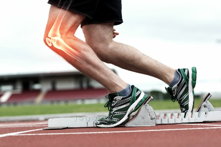
During flexion and extension movements of the knee, the kneecap slides through a guide groove of the femur. However, the kneecap can pop out of this groove due to an unfortunate twisting motion or a lateral impact. If this occurs, it is called a patellar luxation. This can happen during sports and also in everyday life and cause a pain. With the help of your orthopedist, you can get rid of the stress of knee injuries.

What is patellar luxation?
The patella is stabilized by its shape, the guide groove in the femur, and surrounding ligaments and muscles. Due to a lack of muscular guidance, but also as a result of previous patellar injuries, the patella can deviate from the sliding bearing or the guide groove or, in the worst case, pop out.
With patellar luxation, the kneecap slips out of place, causing severe pain as well as a loss of function with every movement. The lateral ligaments may be under too much tension or may tear completely. It is not uncommon for the dislocated patella to slide back on its own in a patellar dislocation, but sometimes it remains in its dislocation site on the outside of the knee joint.
With an incidence of 5.8/100,000 in the normal human population, patellar luxation is one of the most common knee joint injuries.
Causes of knee injuries
There are multiple triggers for patellar luxation. Jumping out can occur in patients of any age, but is often a problem in younger girls or women.
The dislocation forms are divided into acute constitutional, acute traumatic and habitual.
In the acute constitutional form, certain factors are responsible for inadequate trauma being sufficient to cause the patella to dislocate.
The following predisposing factors increase the risk of possible patellar dislocation:
- A congenital malformation of the patella and the sliding bearing (dysplasia).
- An axial or rotational malposition of the knee joint ("knock knees" or genu valgum)
- A raised patella (patella alta) and an external attachment of the patellar tendon to the tibia
- A muscle imbalance
- A change in the retaining ligaments of the kneecap
- Ligament laxity in the course of pathologies such as Ehlers-Danlos syndrome, arachnodactyly, osteogenesis imperfecta, Turner syndrome, trisomy 21, Kabuki syndrome
- In the context of (other) syndromes, such as diastrophic dysplasia, Ellis-van-Creveld syndrome or Scholte syndrome.
In the acute traumatic form of patellar luxation, rotational trauma or direct force to the knee can cause the patella to pop out.
Sports with rapid changes of direction increasethe risk of dislocation.
Other forms of patellar luxation
Another form of patellar luxation is the habitual one with a frequency of about 20% of all luxations. Habitual forms are volitionally triggered and dislocations occur in everyday movement without trauma, usually involving a pre-damaged patellofemoral joint.
In addition to the forms listed, there are other, rarer causes, such as congenital dislocations, which may already be present at birth, neurogenic dislocations, due to an abnormal pull of the thigh muscles, or so-called iatrogenic dislocations, after insufficient operations on the patella or the leg axis.
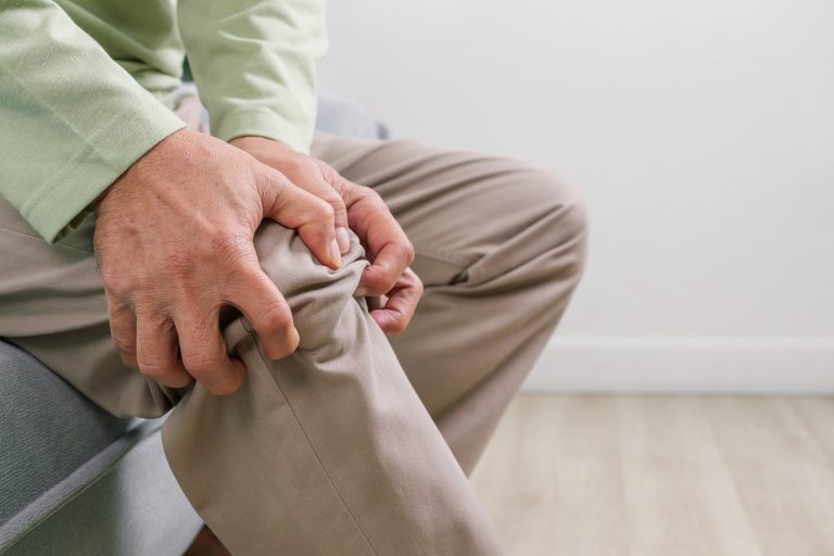
Symptoms
Possible symptoms of patellar dislocation include pain, swelling of the knee, and possible hematoma discoloration. Acute first dislocation is usually very painful, repetitive dislocations usually less so or not at all. In general, the more often the patella luxates, the greater the likelihood of symptoms decreasing in intensity.
Most common course of patellar luxation
Almost always, the kneecap pops out to the outside of the affected knee, usually causing rupture of the joint capsule and the ligament that stabilizes the kneecap on the inside of the knee, called the medial patellofemoral ligament or MPFL for short. During the dislocation itself, a popping sound may be heard and it feels as if the knee is “dislocating”, usually this is also clearly visible by a slipped patella.
Possible injuries and risk factors
In the case of the traumatic knee joint effusion or possible hemarthrosis cited, possible concomitant injuries to the dislocation as the cause of the effusion must be noted and considered. With persistent dislocation, the knee is almost always in a flexed rest position. The mobility of the knee joint may be severely restricted due to pain even after reduction has been completed.
Consequences of patellar luxation
Once the kneecap has popped out, the knee is more unstable after that. This can lead to anterior knee pain, especially during more intense exercise. Longer-term effects of patella dislocation or especially repeated dislocations, which usually also cause more damage to the cartilage, can increase that as risk factors for knee osteoarthritis.
Diagnostics
A luxated patella is usually clearly recognizable and easily visible. Patellar luxation is often a diagnosis at a glance. The malposition of the patella lateral to the knee joint can be seen particularly well in a lateral comparison. In most cases, a clear knee joint effusion, which appears in the form of a so-called “dancing patella”, is palpable.
The apprehension test of the patella can also be used to detect a tendency to dislocation in cases where patellar dislocation has occurred but the patella has spontaneously reduced. However, this should only be performed with great caution, as it can lead to new dislocations after a dislocation has already taken place.
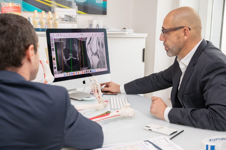
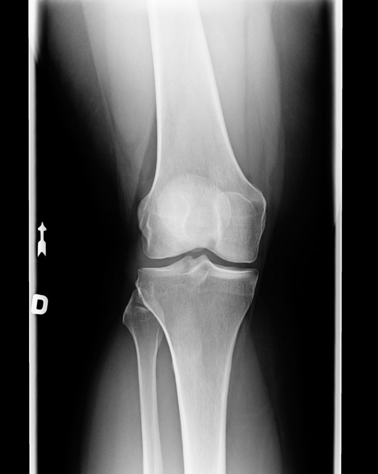
Clarity through X-ray and MRI
Radiographs can be used to rule out associated bony lesions and detect possible predisposing factors. This can be used to assess the shape and position of the patella, as well as the gliding groove.
Magnetic resonance imaging (MRI) is used to evaluate the ligamentous apparatus, as well as bone and cartilage structures, which can also be used to identify a possible rupture of the medial patellofemoral ligament (MPFL) mentioned.
Other possible diagnostic procedures that also provide added therapeutic value are knee joint puncture and arthroscopy.
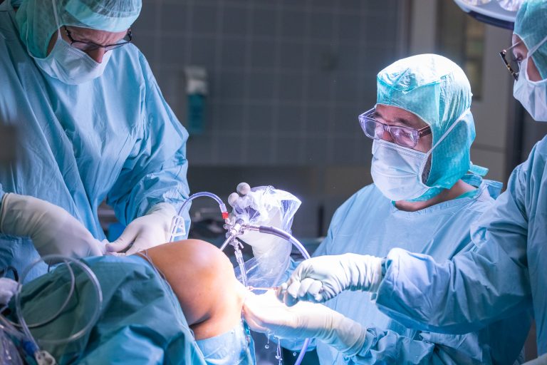
Therapy options
In the case of patellar luxation, a distinction is made between a conservative and a surgical therapy procedure.
Conservative procedure
Conservative therapy involves reduction of the patella and immobilization of the joint with a plaster sleeve. In addition, the affected knee should be elevated and cooled. Forearm crutches, appropriate analgesic medication, and thromboprophylaxis are also among the acute measures.
In the further course, the joint of the knee is fixed or stabilized with a 4-point orthosis with gradual limited flexion and appropriate physiotherapy should be started as soon as possible.

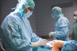
Operative procedure
In the surgical procedure, on the one hand, the medial patellar ligament apparatus and in particular the so-called MPFL (ligamentous connection between the patella and the thigh) can be reconstructed or the knee or its guidance can be realigned. The final treatment and surgery are individualized from case to case, and in some cases several different procedures are combined.
Surgical procedures after knee injury
MPFL reconstruction (medial patellofemoral ligament): In cases of mild malformation of the gliding groove and when the patella is not central, the medial ligament between the patella and the thigh can be sutured, tightened or completely reconstructed. This can prevent a new dislocation from the plain bearing. For this purpose, an autologous tendon of the thigh is used for the reconstruction, which is fixed minimally invasively via small skin incisions on the patella and the thigh.
Tuberosity osteotomy: If the kneecap is too high or too far out, the attachment of the patellar tendon to the tibia can be loosened with a bone flap and brought into the anatomically correct and desired position. The detached bone flake is fixed with two or three screws.
Trochleaplasty: If the gliding groove (trochlea) is poorly formed or completely absent, it can be deepened on the thigh. For this purpose, the knee joint is opened during an operation and the cartilage with a fine layer of bone is detached from the thigh in order to be able to excavate the desired recess. Subsequently, the detached scale is firmly anchored again with sutures.
Adequate aftercare of the knee joint
Regardless of which procedure is ultimately chosen and suitable for the respective patient, adequate aftercare is just as crucial as a good operation. Depending on the procedure, the knee joint is relieved or partially relieved of weight-bearing for a few weeks and then gradually increased until full weight-bearing is achieved.
In addition, muscle strengthening and coordination training are essential as part of postoperative physical therapy. The healed extensor muscles can again assure the necessary support.

Conclusion
Recovery from a patellar dislocation depends on how badly the knee was injured and how it is treated. When and how intensively the knee joint can be exercised and loaded again depends on the personal requirements, the respective type of sport and the success of the treatment.
The ideal and individually best type of therapy depends on the patient, the anatomical conditions and the type of injury.
However, adequate treatment, whether conservative or surgical, is important in order to prevent possible late complications, such as new dislocations, chronic instability or osteoarthritis in the knee joint, as best as possible.
The appropriate treatment should always be done in consultation with your treating orthopedist to achieve the best possible outcome for you.