
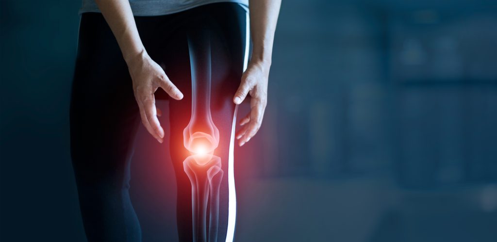
Cartilage damage in the knee | knee arthrosis (gonarthrosis)
Osteoarthritis of the knee is a degenerative disease of the knee joint that often leads to pain and limited mobility. The condition is often the result of years of wear and tear on the joint, an injury or another condition such as rheumatoid arthritis. Cartilage damage in the knee is a common result of this wear and tear. The pain is caused by the grinding together of bone surfaces that are no longer protected by cartilage.
Dr. Martin Gruber is a knee specialist in Vienna and specializes in the treatment of osteoarthritis and cartilage damage in the knee, and thus also in the treatment of knee pain. Treatments can help relieve symptoms, slow disease progression, prevent further complications and improve your quality of life with knee cartilage damage. Learn more about this clinical picture, its causes and possible forms of therapy below.
OVERVIEW
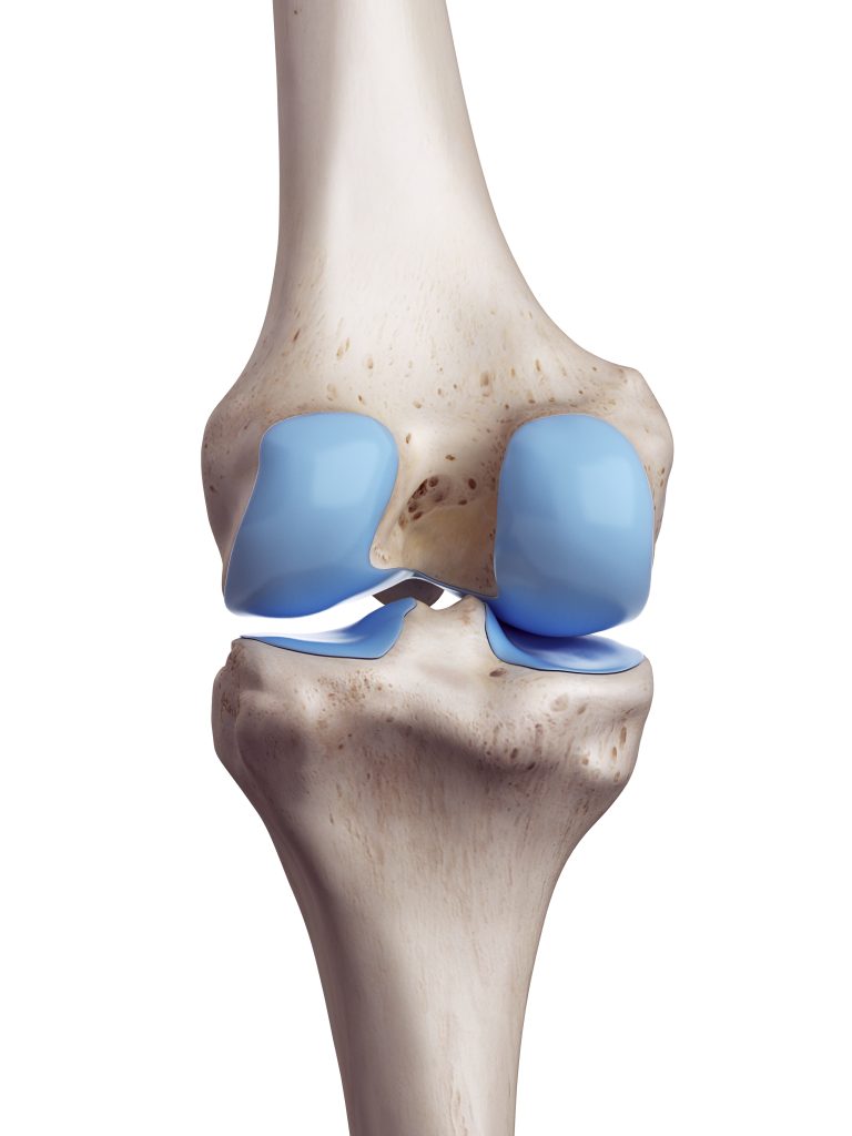
What is cartilage?
Articular cartilage is the cartilage tissue that covers the articular surfaces of all true joints. Hyaline cartilage is firmly attached to the underlying bone and is the gliding surface that makes painless and frictionless movement in a joint possible in the first place. The cartilage layer is very elastic to pressure and distributes the pressure in the joint as evenly as possible like a shock absorber.
How does cartilage damage occur in the knee?
Cartilage damage in the knee can occur acutely as a result of an accident, but also as a result of chronic misuse or overuse, as well as inflammation and circulatory disorders.
In acute, traumatologically induced cartilage injuries, the sudden pressure and shear forces cause localized cartilage damage, usually in the main loading zone. Possible causes of chronic cartilage damage include malalignment of the leg axis (knock knees or bow legs), obesity, meniscus damage, or instability in the knee joint. As a result of incorrect or excessive loading, the cartilage loses elasticity and thus its “shock absorption” and, as it progresses, increasingly loses its substance and function.
Prerequisites for the insertion of a knee prosthesis
Cartilage damage is classified according to the severity of the cartilage injury or degeneration. The newer, somewhat differentiated classification according to ICRS (International Cartilage Research Society) subdivides:
- Grade 0: Normal findings with smooth, stable cartilage, no discernible defects
- Grade 1: cartilage softening with intact surface and/or superficial cracks or fissures in the cartilage
- Grade 2: Tears with a depth < 50% of the total cartilage thickness (abnormal cartilage).
- Grade 3: cartilage defects with a depth > 50% of the total cartilage thickness, i.e. possibly reaching the bone layer
- Grade 4: complete cartilage lesion, i.e. the bone under the cartilage is exposed ("cartilage baldness")
What is the procedure for artificial knee joint surgery?
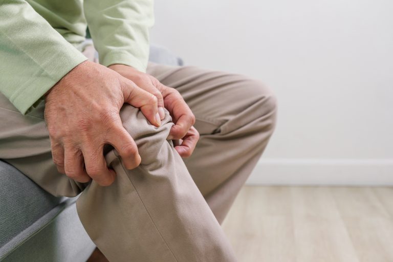
If the cartilage is injured or worn, it is often recognized by a crunching, creaking or cracking sound in the joint; pain and chronic inflammation are the result of insulted cartilage. However, unlike bone, articular cartilage itself has neither a blood nor a nerve supply. For this reason, the pain does not originate from the cartilage itself, but from the underlying bone and joint capsule.
The pain often occurs when walking down stairs or hills, as well as after exertion; a start-up pain when standing up after prolonged sitting is also typical. The degree of discomfort depends on the size and depth of the defect, as well as its localization. Chronic cartilage damage often leads to pain on start-up, strain and inflammation. In addition, patients report swollen knees and limited mobility.
Acute injuries with cartilage damage result in sudden discomfort and joint swelling. Pieces of cartilage that can become completely detached during an injury can lead to a blockage in the knee joint.
What are the risks and side effects of knee replacement?
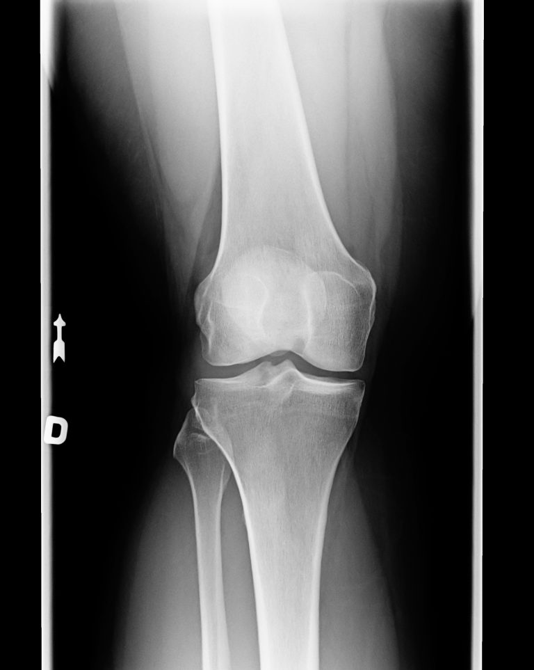
A precise survey of the pain symptoms, as well as a thorough clinical examination, can provide initial indications of possible cartilage damage in the knee joint. An X-ray examination of the affected knee is mainly used to exclude a bony injury, but may already indicate advanced cartilage damage or arthrosis if there are radiological signs of wear.
The diagnosis of cartilage damage is confirmed by magnetic resonance imaging (MRI), which can be used to determine the size and depth as well as the exact location of the cartilage damage. In addition, an X-ray examination of the leg axis should be performed if a malposition (knock knees or bow legs) is suspected, as this can often be the cause of the cartilage damage. In the case of cartilage damage to the kneecap, instability of the kneecap should also be investigated.
What do I have to take into account after the operation of an artificial knee joint?
Depending on the depth, size and location of the cartilage damage, there are different treatment methods. Superficial and smaller cartilage defects can sometimes be treated conservatively(physiotherapy, infiltrations , or medicinal pain therapy); large and deep damage should be repaired surgically.
Conservative therapy
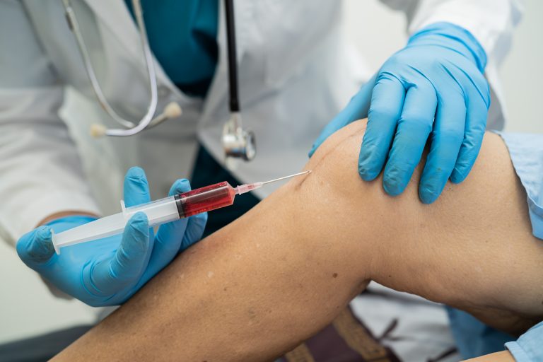
In addition to the aforementioned physiotherapy and medicinal pain therapy, nutritional supplements containing ingredients of articular cartilage and, above all, infiltrations using hyaluronic acid can help to prevent further wear and tear as well as a progression of the cartilage damage. The infiltrations allow the cartilage to store more hyaluronic acid and the synovial fluid to better perform its function as synovial fluid. Various preparations are available for this purpose.
Cartilage smoothing
This procedure is often performed carelessly, because a positive effect is not scientifically proven. Cartilage smoothing should therefore not be performed as a stand-alone procedure. In this procedure, frayed and lifted cartilage is removed during arthroscopy.

Microfracturing
At the site of the injury, the cartilage is cleanly removed and the now exposed bone is finely punctured with small holes. In this way, stem cells from the bone marrow enter the exposed area and recreate a tissue very similar to cartilage. However, this surgery does not work for injuries that are too large and requires intact bone beneath the cartilage defect. The decisive factor is the follow-up treatment: relief with crutches and physiotherapy for at least six weeks.
OATS (Osteochondral Autologous Transplantation or Cartilage Bone Grafting).
In this procedure, a cartilage-bone cylinder is harvested from an area of the cartilage that is not under stress and inserted into the injured or worn area. The removed broken part is reinserted at the removal site – so the cylinders are simply swapped and the cartilage is restored to full function where it needs to be able to bear weight. In contrast to microfracturing, this method can be used to treat deep defects that extend into the bone, but limitations include the availability of bone-cartilage cylinders, as well as the potential pain that can occur at the harvest site. When several cylinders are used, it is called a mosaicplasty, which is particularly suitable for the treatment of Ahlbäck’s disease (aseptic bone necrosis), but also for OCD (osteochondrosis dissecans).
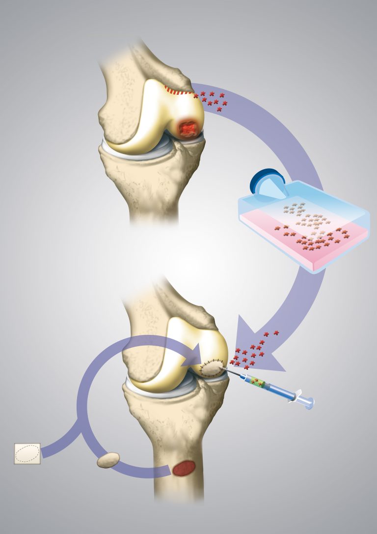
ACT and MACHT (cartilage cell transplantation)
Cartilage cells are removed and cultivated in the laboratory until sufficient new “endogenous” cartilage tissue has grown back. The method is particularly suitable for large, well-limited cartilage damage. This is then reinserted into the knee. The problem with this is that it requires two surgeries on the knee, one procedure to remove tiny pieces of cartilage from an unstressed area of the knee joint and a second procedure in which the finished cartilage cell graft is inserted into the cartilage defect. A simpler form of therapy is cartilage coverage with the AutoCart System (Minced Cartilage) from the company Arthrex, in which even larger cartilage damage can be repaired in usually one minimally invasive procedure. Stem cell transplantation offers another way to avoid a second surgery.
Stem cell transplantation
Stem cells are removed from the bone marrow of the pelvic bone in an operation, separated and immediately inserted into the cartilage. The advantage: there is no waiting time of up to six weeks between removal and insertion.
Conclusion
Cartilage damage in the knee joint can occur either at the kneecap or at the joint surfaces. A distinction is made between acute damage, which is usually caused by accidents or injuries, and chronic damage, which is usually caused by chronic overuse or overload. Depending on the cause and the extent of the cartilage defect, a distinction is made between two therapeutic approaches. An approach that eliminates the cause and therapy directly at the knee joint.
This can be done conservatively, but also surgically. Particularly in the case of surgical cartilage rehabilitation, the choice of the respective surgical method is of great importance, along with consistent follow-up treatment.
Your treating orthopedist will help you find the appropriate form of therapy and achieve the best possible clinical outcome. Contact me!